
Pneumonia
last authored: Feb 2010, David LaPierre
last reviewed:
Introduction
Pneumonia, or infection of the lower respiratory tract, is one of the most important causes of death, especially during adult years. It can be caused by a number of pathogens, including bacteria, viruses, fungi, protozoans, and parasites.
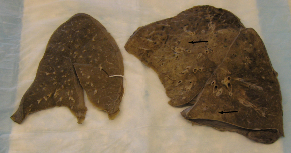
normal lung on left; bronchopneumonia (arrows) on right.
courtesy of Dr Zhaolin Xu, Department of Pathology, Dalhousie University
As it is often readily curable, every health care provider should be on the lookout for it, and every physician should know how to rapidly diagnose and treat pneumonia.
The Case of Henry C
Henry Chu is a...
Causes and Risk Factors
Risk factors for pneumonia include:
- smoking
- chronic lung diseases
- alcohol, drug use (risk of aspiration)
- neurological diseases (stroke, ALS, etc)
- toxic inhalation
- mechanical obstruction
- intubation
- immunosuppression, splenectomy
- most common pathogens
- bacteria
- viruses
most common pathogens
The following pathogens represent the most common causes of pneumonia in different populations:
community-acquired in healthy adults |
|
community-acquired in elderly/ |
|
nosocomial |
|
HIV-associated |
|
alcoholic |
|
ventilator-associated |
|
immunocompromised |
|
Pathophysiology
Under normal circumstances, the lungs are sterile. Pathogens can enter through inhalation, by hematogenous spread, from a contiguous focus of infection, or most commonly by aspiration of oropharyngeal secretions (almost 50% of healthy men aspirate some oropharyngeal contents during sleep). Infection can also follow inhalation of aerosolized droplets, as occurs with tuberculosis.
Defenses against pulmonary colonization include cough, mucociliary clearance, and other innate immune defenses. Impaired normal defense mechanisms frequently contribute to the development of pneumonia. These can include:
- reduced cough reflexes (drugs, alcohol, neuromuscular disease)
- impaired ciliary transport (smoking, cystic firbosis, COPD)
- chronic diseases
- immunologic deficiency
Infection can spread along airways and through alveolar walls, and exudative inflammation results.
Lobar Pneumonia:
- congestion from day 0-2 - exudate
- red hepatization days 2-4 - appears like liver as consolidation occurs
- gray hepatization days 4-8 - less congestion
- resolution
History and Physical Exam
A thorough history and physical can help diagnose pneumonia and also distinguish among different pathogens.
- history
- physical exam
History
Respiratory symptoms of pneumonia include:
- cough
- persistent, hacking - Mycoplasma
- sputum production
- dyspnea (shortness of breath)
- chest pain, especially pleuritic
- fever
- malaise
- absence of rhinorrhea or sore throat
A history of rhinitis or pharyngitis suggests respiratory virus, Mycoplasma, or Chlamydia. Abrupt onset of myalgia, arthralgia, headache, and fever are commonly seen in influenza virus infection.
Abrupt onset of myalgias, arthralgia, headache, and fever suggest influenza or mycoplasma.
Duration of symptoms is a critical piece of information:
- pneumococcal, mycoplasma, or virual or viral disease is usually acute, with symptoms lasting for hours or days.
- Symptoms lasting 10 days or longer are rarely caused by common bacterial pathogens and should raise suspicion of mycobacterium, fungal, or anaerobic pneumonia, and/or the presence of underlying disease.
Review of symptoms
- diarrhea - suggests Legionella
- altered mental status - ?meningitis
past medical history
- asthma
- dementia
- immunosupression
Investigations
- lab investigations
- diagnostic imaging
Lab Investigations
blood tests
- CBCD: usual elevated white cell count, with left shift; higher for bacterial infection
- lytes
- urea, sCR
- troponin/CK
- AST, ALT, ALP,
- bili: modest increases in conjugated bilirubin are seen in many bacterial infections
- C-reactive protein: monitoring severity of infection and treatment response
- blood culture: will be positive for 20-30% of people with bacterial pneumonia.
nasopharyngeal swab: influenza, parainfluenza, metapneumovirus, RSV, adenovirus
sputum smear and culture: must be done carefully to avoid contamination by oropharyngeal flora. Both false negatives and false positives are common and should be interpreted carefully.
- gram stains
- Gomori's Methenamine Silver (GMS stain)
- PAS/diastase stain
- Ziehl-Neelsen stain for mycobacteria: should be done unless diagnosis of acute bacterial pneumonia is clear
- viral inclusions
other tests:
- urinalysis
- tuberculin skin test
- lumbar puncture should be done in patients with penumonia and confusion to rule out meningitis
- mycoplasma IgM
- tissue biopsy can be very helpful, but its invasiveness is an important consideration.
Differential Diagnosis
The differential diagnosis of pneumonia includes:
- heart failure
Treatments
- immediate
- community-acquired
- nosocomial
- nursing home
Immediate Treatments
As with any patient, it is important to begin by treating the vitals, or ABCs, of the patient. This includes, as necessary:
- oxygen administration and monitoring of saturation
- positioning
- IV access
- salbutamol/Ventolin
For hospitalized patients, length of stay has been shown (Carratala et al, 2012) to be minimized by a clinical pathway prompting:
- early mobilization
- switch from IV to oral antibiotics when clinical improvement was seen, and when vital signs were normal
- discharge home when on oral antibiotics, when cognition was improved, and when oxygen status was at baseline
Consequences and Course
Complications of pneumonia include:
- acute respiratory distress syndrome (ARDS)
- pleural effusion/empyema (infection in the thoracic cavity)
- lung abscess - destruction of parynchema and collection of pus
- pleuritis
- purulent pericarditis
- hyponatremia
- sepsis
- brain abscess and meningitis via hematogenous spread
- septic arthritis
- death
Prognosis can be predicted with scoring tools, such as the Pneumonia Severity Index (PSI) and the CURB-65 and CRB-65. All three appear to be similarly accurate (Ochoa-Gondar O et al, 2011).
Increased mortality occurs in patients with:
- cardiac or pulmonary disease
- cirrhosis
- elderly
- leukopenia, leukocytosis, hypoxemia, bacteremia
Resources and References
Carratala J et al. 2012. Effect of a 3-Step Critical Pathway to Reduce Duration of Intravenous Antibiotic Therapy and Length of Stay in Community-Acquired Pneumonia: A Randomized Controlled TrialA 3-Step Critical Pathway for CAP. Arch Intern Med. 172(12):922-8.
Harris J et al. 2010. British Thoracic Society guidelines for the management of community acquired pneumonia in children: update 2011. Thorax. 66 Suppl 2:ii1-23.
Jung YJ, et al. 2010. Effect of vancomycin plus rifampicin in the treatment of nosocomial methicillin-resistant Staphylococcus aureus pneumonia. Crit Care Med ;38(1):175-180.
Meijvis SC et al. 2011. Dexamethasone and length of hospital stay in patients with community-acquired pneumonia. Lancet 377(9782):2023-2030.
Nazarian et al. 2009. Clinical policy: Critical issues in the management of adult patients presenting to the emergency department with community-acquired pneumonia. Ann Emerg Med. 54(5):704-731.
Ochoa-Gondar O et al. 2011. Comparison of three predictive rules for assessing severity in elderly patients with CAP. Int J Clin Pract. 65(11):1165-72.
 |