
Electrocardiogram (ECG) Placement
last authored: March 2012, David LaPierre
last reviewed: March 2012, Stacie Vriesema
Introduction
For information on ECG background and interpretation, see here.
The electrocardiogram (ECG) represents electrical activity originating in the heart and captured on the skin. It is of profound importance in assessing cardiovascular disease and other conditions affecting heart function.
The standard 12 lead ECG has 6 limb leads (I, II, III, avR, aVL, and aVF) and six precordial (chest) leads (V1-V6).
Patient Procedure
Place the patient supine, with the head slightly elevated. Ensure that the arms and legs are supported from underneath.
Expose the chest and limb areas. Where necessary, shave the skin to allow secure placement of the leads.
Ask the patient to remain still, with their muscles relaxed, and to breathe normally.
Once the leads are placed, enter the patient's age as prompted, and press the '12 lead' button.
Limb leads
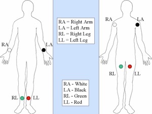
two suggested limb lead placements, from wikipedia
The limb leads are composed of standard leads I, II, and III and augmented leads aVL, aVR, and aVF.
Place the electrodes as follows:
- LA: left arm - either on the shoulder, or the forearm
- RA: right arm - as above
- LL: left leg - either just below the knee, or on the hip
- RL: right leg - as above
Augmented leads are termed unipolar because they use one positive electrode as reference against a combination of the other limb electrodes. aVL uses the left arm as a positive electrode, aVR uses the right arm, and aVF the left leg.
Precordial Leads
The precordial leads are placed on the chest, just above the heart. Their positions are as follows:
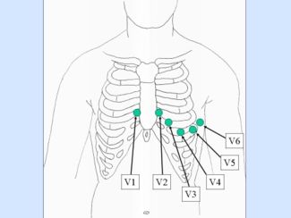
from wikipedia
V1: fourth intercostal space, at right edge of sternum (red)
- feel for the space between the clavicle and the rib beneath, place one finger there and feel down with subsequent fingers until your last finger is resting on the 4th intercostal space
V2: fourth intercostal space, at left edge of sternum (yellow)
V3: midway between V2 and V4 (green)
V4: fifth intercostal space, midclavicular line (blue)
V5: same horizontal line as V4, left anterior axillary line (brown)
V6: same horizontal line as V4, left mid axillary line (purple)
remembering the colours
a stop light goes red, yellow, green; blue skies in spring,
brown leaves in autumn, and a purple Christmas!
(cortesy of Pauline Chordash)
When placing leads on females, or men with generous chest tissue, lift up the breast to place the lead on the chest wall; do not place it on the breast.
15 and 18 Lead ECG
15 or 18 lead ECGs can be done with alternate precordial lead placement to assess for posterior- or right-sided disease. While the 18-lead ECG is perhaps more sensitive for early detection of ischemia or infarction, in practice, either should be used for:
- all inferior infarcts
- suspected right ventricular or posterior infarcts: ST depression in V1 and V2, with R waves
- continous chest pain and a non-diagnostic 12 lead ECG
15 lead ECG requires repositioning of the leads as follows:
Ensure the ECG is relabelled as a 15 lead. |
18 lead ECG requires repositioning of the leads as follows:
Again, ensure the ECG is relabelled as an 18 lead ECG. |
Resources and References
http://sprojects.mmi.mcgill.ca/heart/egcyhome.html
ECG simulation and explanation
 |