
Cardiac Arrest
last authored: Oct 2014, David LaPierre
last reviewed: Nov 2014, Shannon Masse
Introduction
Cardiac arrest, also called cardiopulmonary arrest or circulatory arrest, is the cessation of normal circulation of the blood due to failure of the heart to contract effectively.
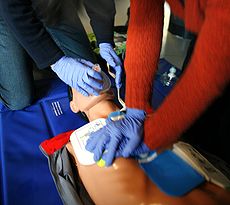
CPR; used with permission
Without timely CPR and defibrillation, cardiac arrest signifies death. Cardiac arrest can occur without warning due to a number of reasons, but in many patients, it is the expected, final outcome to a serious illness.
When in the hospital, the response to cardiac arrest is often termed a 'code' or 'code blue'.
The cardiac rhythms accompanying cardiac arrest may be called 'shockable' and 'non-shockable', depending on their potential responsiveness to defibrillation:
shockable rhythms
|
non-shockable rhythms
|
The Case of Will R.
Will R. is a 78 year-old man who suffers cardiac arrest at the mall. CPR is started within seconds, and he is unsuccessfully resuscitated with the mall's AED. An ambulance crew brings him into the emergency department, intubated and with an IV in place, with his wife beside him.
As team leader, what do you do?
Causes and Risk Factors
The majority of cardiac arrests are caused by acute coronary syndrome, or myocardial infarction (heart attack).
Other cardiac conditions that may lead to cardiac arrest include:
7 H's and 5 T's: pnemonic for mechanisms
- hypoxia
- hypovolemia
- hyperkalemia
- hypokalemia
- hypoglycemia
- hypothermia
- hydrogen ions (acidosis)
- thrombosis (MI, PE)
- tension pneumothorax
- tamponade
- toxins/therapeutics
- trauma
- arrhythmias, both brady- and tachy-
- genetic abnormalities, including Wolff-Parkinson-White syndrome, long QT syndrome, and Brugada syndrome
- congestive heart failure
- dilated or hypertrophic cardiomyopathy
- congenital structural or arterial abnormalities
- valvular disease, especially aortic valve stenosis, mitral valve stenosis
- myocarditis
- cardiac tamponade
- pulmonary hypertension
Non–cardiac causes of sudden cardiac death include:
- hyperkalemia
- hypokalemia
- pulmonary embolism
- acidosis (various causes)
- blunt or penetrating trauma to the chest
- non-trauma related bleeding (such as gastrointestinal bleeding, aortic rupture, and intracranial hemorrhage)
- overdose
- drowning
Pathophysiology
main article: electrical control of the heart
Ventricular fibrillation (VF) represents the loss of coordinated contraction of the ventricles. It is often preceded by ventricular tachycardia (VT). As the heart function becomes increasingly unstable, reentry circuits occur. These reentry circuits, with conduction occurring outside the regular cardiac cycle, are thought to break up into smaller wavelets. These impulses wander throughout the ventricles, culminating in VF. Both VF and VT may be caused by the factors listed above.
A rapid loss of cardiac output will result asystole and death if VF or pulseless VT is not quickly reversed by defibrillation.
Pulseless electrical activity (PEA) is a group of rhythms that are organized or semi-organized, but lack a discernible pulse. Examples include:
- idioventricular rhythms
- ventricular escape rhythms
- postdefibrillation idioventricular rhythms
- bradysystolic rhythms
Signs and Symptoms
- history
- physical exam
History
Due to inadequate cerebral perfusion, the patient will be unconscious and will have stopped breathing.
If possible, rapid efforts should be made to understand the patient’s symptoms prior to the event, as well as their health history.
Attempt to clarify code status to determine whether resuscitation should be attempted. If this information is unavailable, attempt resuscitation.
Sudden cardiac arrest may occur due to channelopathy. Family history should be probed for events such as sudden death, syncope, seizure, or drowning.
Physical Exam
Cardiac arrest results in a lack of circulation. This may be demonstrated physically by:
- nonpalpable pulse - radial, or carotid
- lack of heart sounds
- evidence of non-perfusion - poor colour, no movement or agonal movement
- apnea or agonal breathing
However, an absent pulse can sometimes be difficult to identify with certainty. If suspicion is high, and resuscitation is warranted, emergency efforts should be started without delay.
Investigations
- lab investigations
- diagnostic imaging
Lab Investigations
Bloodwork to be drawn in addressing cardiac arrest can include:
- glucose (fingerpick)
- electrolytes, especially potassium
- troponin
- toxicology screen
Diagnostic Imaging
Beyond cardiac monitoring, there are few indications for imaging during cardiac arrest. Some clinicians may use bedside ultrasound to assess for tamponade, pneumothorax, or cardiac activity.
If resuscitation has been successful, ECG, chest X-ray, and other modalities may be used to better understand the cause.
Treatments
The interval from collapse to defibrillation, the only effective treatment for VF, is one of the most critical determinants of survival. CPR cannot directly restore an organized rhythm, but does provide a small amount of blood flow to the brain and heart. For every minute that passes, survival declines by 7-10% per minute, or by 3-4% per minute if CPR is provided (AHA 2010 guidelines).
Levels 1 and 2: First Responder and Primary Care Clinic.
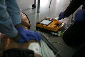
CPR with AED, used with permission
First responders should provide immediate basic life support (BLS), including early defibrillation with an AED if available. Call for help to bring other needed equipment and organize transport.
Level 3: Community Hospital
Resuscitation within the hospital setting should follow the protocols of advanced cardiac life support (ACLS), centred on highly effective CPR and incorporating defibrillation, IV access, medication administration, advanced airways, and identification of addressable causes of the arrest.
Unfortunately, cardiac arrest frequently ends in death, and providers should be versed in elements of breaking bad news while discussing with family.
Ethical Issues
There are a number of ethical issues surrounding cardiac arrest and resuscitation.
Advance care directives are legal documents that provide directions about a person’s wishes or authorize a surrogate decision-maker to make decisions on behalf of an individual that is unable to do so. Frequently, these directives will include a request that resuscitation should not be attempted. It is important that everyone, especially those who are elderly, have serious medical disease, or are hospitalized, consider whether they would like to receive attempted resuscitation after cardiac arrest. If the patient’s wishes are unknown, it is appropriate to begin resuscitation and make further decisions after consultation with the patient’s relatives or friends.
Resuscitation should not be attempted if there are objective signs of irreversible death (e.g. decapitation, rigor mortis, or decomposition) or if the patient has declined resuscitation. Providing only a token attempt at resuscitation (“slow code”) is not appropriate, as doing so undermines the provider-patient relationship and compromises the ethical integrity of health care providers. If resuscitation is indicated, a full attempt at resuscitation should be made.
Resuscitation efforts may be terminated if, in the opinion of the physician, the patient will not respond to further resuscitative measures.
Consequences and Course
Without immediate resuscitation, and in many cases even with attempted resuscitation, cardiac arrest will rapidly lead to irreversible brain damage and brain death due to a lack of perfusion.
There are at several different measures of successful resuscitation. Return of spontaneous circulation (ROSC) is the return of a sustained, palpable pulse in a patient who previously was pulseless. Cardiac arrest survivors do not always survive without neurological deficits, with good quality of life, and this is another important indicator.
Rates vary for out-of-hospital and in-hospital arrests. One large study done in Australia for out-of-hospital arrests showed a survival to hospital discharge rate of 13% of attempted resuscitations. Of these, 8% died within a year; with those that lived beyond a year, 55% were assessed to have a ‘good’ recovery (Smith et al, 2014).
Regarding in-hospital arrests, a large study done in hospitals across the US showed that in 2009, 54% of acute resuscitation was successful, 22% of patients survived to discharge, and that rates of neurological disability were 28% (Girotra et al, 2012).
Resources and References
Girotra et al. 2012. Trends in Survival After In-Hospital Cardiac Arrest. N Engl J Med. 367(20):1912-20.
Smith K, Andrew E, et al. (2014). Quality of Life and Functional Outcomes 12 Months After Out-of-Hospital Cardiac Arrest. Circulation. Oct 29. pii: CIRCULATIONAHA.114.011200.
Morrison et al. 2010. 2010 American Heart Association Guidelines for Cardiopulmonary Resuscitation and Emergency Cardiovascular Care Part 3: Ethics. Circulation. 122: S665-S675.
Topic Development
authors: Brandon Cook, David LaPierre
reviewers: Shannon Maase
 |