
The Placenta
last authored:
last reviewed:
Introduction
The placenta acts as the interface between the fetus and mother. It is referred to as a fetomaternal organ as it is composed of tissues from both beings.
During the embryonic period, the adnexa includes the amnion, chorion, umbilical vesicle (yolk sac) allantois, and connecting stalk.
The chorion
The placenta develops in a highly dynamic process. It appears first as the chorionic sac in week two, which surrounds the embryo and consists of two layers - an outer ectoderm (trophoblast) layer and inner mesoderm. The trophoblast, composed of cytotrophoblast and syncytiotrophoblast, is responsible for implantation and invasion into the endometrium during the first week.
Chorionic Villi
Primary chorionic villi appear at the end of the second week from cytotrophoblasts, and mesenchyme begins to invade early in the third week to form secondary villi. Some mesenchymal cells soon differentiate into capillaries and blood vessels, creating tertiary villi. Arteriocapillary networks form and connect to the embryonic circulation through the connecting stalk and chorion. By the end of the third week, embryonic blood is carrying oxygen and nutrients to the embryo.
At the same time, cytotrophoblasts of the chorionic villi proliferate and form the cytotrophoblastic shell around the synthiotropblast, gradually forming the bridge between the chorion and the endometrium. Stem chorionic villi attach to maternal tissues, and from these branch chorionic villi grow and provide the main exchange of material.
The Decidua
The decidua is the component of the endometrium that falls away from the uterus after birth. THere are three regions:
- decidua basalis: contributed by the mother
- decidua capsularis: surrounds chorion
- decidua parietalis: covers remainder of endometrium
Intervillous space, present between the chorionic villi and decidua basalis, is a large blood-filled space. Maternal aretries and veins penetrate the cytotrophoblastic shell.
As the amniotic sac enlarges faster than the chorionic sac, they soon fuse to produce the amniochorionic membrane. It in turns adheres to the decidua capsularis and, following the capsular disappearance, becomes fused with the decidua parietalis.
Amnion and Amniotic Fluid
The amnionic sac develops from extra-embryonic ectoderm, being attached to the margins of the embryonic disc. Following embryonic folding, its junciton with the embryo occurs on the ventral surface. As it expands, it gradually takes over the chorionic cavity and the epithelial covering of the umbilical cord.
The volume of amniotic fluid increases slowly, from 30 ml at 10 weeks to almost 1000 ml by 37 weeks. Large amounts of fluid are absorbed by the mother, but the fetus also swallows up to 400 ml of fluid per day, absorbing it and returning the fluid to the mother through the umbilical cord. Stenosis or atresia of the gut can lead to hydramnios, or a high volume of amniotic fluid.
Amniotic fluid performs various functions:
- acts as barrier to infection
- cushions embryo
- permits normal lung development
- enables the fetus to develop and move freely
- maintains fluid and electrolyte homeostasis
Amniotic fluid can be examined. High levels of AFP suggest severe neural tube defect, while low levels are suggestive of chromosomal aberrations such as Down syndrome.
Amniotic fluid contains stem cells capable of differentiation into each stem cell lineage. (Coppi et al, 2007)
Umbilical Cord
the umbilical cord consists of an umbilical vein which carries oxygenated blood, and 2 umbilical arteries that return deoxygenated blood (in a normal 3 vessel cord). The umbilical vein is thin walled and especially susceptible to compression, during cord prolapse the vein is compressed at lower pressures. At higher pressures the arteries will also be compressed. The decreased in blood flow triggers baroreceptors causing vagal stimulation which causes decelerations (lower heart rate). Chemoreceptors can sense hypoxemia and thus may raise the baseline heart rate to a tachycardic range.
These vessels are buried within Wharton's jelly. The umbilical arteries are branches of the internal iliac arteries. The umbilical vein flows into the hepatic system, both portal to the liver and the hepatic vein into the IVC. The exchange of oxygen and nutrients between the fetal and maternal circulation occurs via the placenta.
Placental Transport
Infectious agents
- CMV
- rubella
- coxasackie virus
- varicella
- measles
- polio
- Treponema pallidum
- Toxoplasma gondii
Mature Placenta
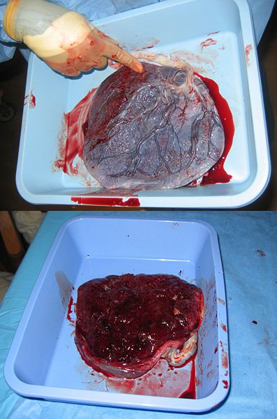
baby side (top) and mother side (bottom)
Resources and References
 |