
Auditory System
last authored:
last reviewed:
Introduction
Hearing
Normal hearing is above ~ 25 dB.
Sound detection, sound localization, speech discrimination, music appreciation
Sound can be transduced through air and bone
Can extract sound you want to hear from the background noise of the world.
The ear actually deconstructs noise
Outer Ear
The prominent pinna and the tragus help collect and focus sound waves into the ear.
The external auditory canal is approximately 2.5 cm long and ends at the eardrum (tympanic membrane).
Middle Ear
The middle ear is an air-filled chamber, with the eardum on one side and the oval window on the other. The eustachain tube connects the middle ear to the nasopharynx.
The middle ear also contains a lever system that takes signals from the tympanic membrane and transfers them to the oval window. The smallest bones in the body - the malleus, incus, and stapes - accomplish this.
The middle ear transfers sound waves from air into the water (perilymph) of the inner ear. This is difficult due to the different impedences, or resistance, of air and water to sound waves. Impedence matching is accomplished by the fact that the tympanic membrane is 20x larger than the oval window, and the lever action of the ossicles. In this way, most of the vibrational energy is transferred to the oval window instead of being reflected.

Two tiny muscles, the tensor tympani and the stapedius, serve to dampen sound waves transferred to the inner ear. They are innervated by the trigeminal and facial nerves.
Inner Ear
The cochlea is a 3.5 cm long tube coiled about 2.5 times into the size of a pea.
It contains several long membranes which vibrate in specific regions according to the frequency of sound - a concept called place coding.
The scala vestibuli is connected to the oval window and is continuous with the scala tympani, which ends in the round window in the inner ear. The scala vestibuli and tympani are filled with perilymph.
The scala media is a separate tube filled with perilymph and contains the hair cells, resting on the basliar membrane.


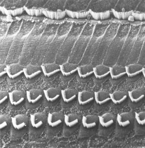
The cochlea contains 4 rows of hair cells - one row of about 3500 inner hair cells and three rows of about 20,000 outer hair cells. Hair cell cilia project into the gelatinous tectorial membrane which lies above them. The tectorial membrane is firmly attached along one edge and hinges up and down.
Presusre waves hitting the oval window are transmitted through the scala vestibuli and in turn through the scala media and scala tympani. The involvement of the basilar membrane, on which the hair cells rest, causes cilia movement and signal tranduction.
The tapering basilar membrane, and its increasing stiffness, results in the auditory place code. Low frequency sounds generate maximum signals at the cochlear apex, while high frequency sounds induce basilar membrane vibration primarily at the base.
About 95% of the 30,000 afferent axons synapse with the few inner hair cells, as the abundant outer hair cells in the organ of Corti instead are involved in sharpening the tuning of the cochlea. Outer hair cells are regulated by sounds waves and by efferent axons leaving the superior olivary complex in the brainstem, responding by shortening their cell body and in some way mediating inner hair cell firing.
outer hair cells dance according to music (youtube video)
Auditory Processing and Pathways
Afferent signaling
Cell bodies of bipolar neurons carrying afferent signals from the cochlea lie in the spiral ganglion. The cochlear nerve joins the vestibular nerve, forming CN VIII. Auditory axons then enter two auditory nuclei (dorsal and ventral) in the brainstem at the cerebellar pontine angle between pons, medulla, and cerebellum.
Brainstem nuclei
The dorsal cochlear nucleus extends to the dorsal acoustic stria and the intermediate acoustic stria.
Pitch analysis
The dorsal cochlear nucleus projects axons that cross the midline and enter the lateral lemniscus, the major ascending auditory pathway. Virtually all fibres terminate in the inferior colliculus in the midbrain, which then sends signals through the brachium to the medial geniculate nucleus, in the thalamus. This is the final relay station before projections reach the auditory cortex on the transverse temporal gyri, mostly buried in the lateral sulcus of the temporal lobes.
Sound localization
Some signals from the ventral cochlear nucleus join the lateral lemniscus and extend to the inferior colliculus, but a greater number end at the superior olivary nucleus of each side, which compare sound intensity (head shadow effect) and the time taken for signals to arrive (interaural delay). The crossing of cochlear nuceli to olivary nuclei occurs in the trapezoid body of the pons. Superior olivary nuclei then project to the ipsilateral infeiror colliculus.
As such, central processing structures receive input from both sides. As such, a central lesion will result in dimunition of signals, but not total deafness.
Efferent signals
The olive sends efferent signals back along CN VIII to the organ of Corti, where outer hair cells are innervated to modulate the sensitivity of the cochlea.
Cortical processing
Tonotopic mapping is maintained all the way to primary auditory cortex, which is located within the Sylvian Fissue of the temporal lobe.
higher auditory pathways process simple cues to:
- sound localization: amplitude cues, temporal cues
- distinguish background noise
- fill in missing bits of information
- detect patterns of sound involved in speech
Problems with Hearing
Resources and References
 |